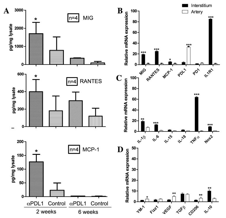
Figure 6.
Treatment with blocking antibodies to PDL1 resulted in increased expression of MIG, RANTES and MCP-1 in allografts by ELISA that was greater at 2 weeks than 6 weeks (A). These samples were taken from the apex of the hearts which contains few large arteries. Microdissection of allografts at 2 weeks demonstrated levels of MIG, RANTES, MCP-1, IL-1R1, IL-1β, IL-6 TNFa and Nos2 were greater in the interstitium than the arterial compartment (B, C). M2 macrophage markers were changed to a lesser extent (D). Bars represent average of 3-4 samples in each group. PCR results represent fold changes compared to allografts treated with control antibody. Differences between interstitial and arterial values were significant at the P<0.05*; <0.01**; or <0.001*** level as indicated.

