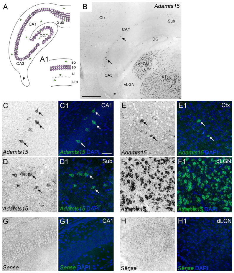
Figure 1.
Sparse expression of Adamts15 mRNA in hippocampus and neocortex. A. Schematic representation of the mouse hippocampus. Excitatory neurons are depicted in magenta. Inhibitory neurons are depicted in green. A1 depicts a higher magnification view of the 4 distinct layers in the CA1 region of hippocampus. B. Localization of Adamts15 mRNA by in situ hybridization in coronal sections of P14 mouse hippocampus. C–F. Shows high magnification images of Adamts15 mRNA in CA1 (C), subiculum (D), layer V of neocortex (E), and dLGN (F) in P14 mouse brain. Cell nuclei are labeled by DAPI staining. Arrows indicate Adamts15 mRNA expression by sparse populations of cells in CA1, Sub, and Ctx. G,H. Shows high magnification images of in situ hybridization with sense control probes in coronal sections of P14 mouse hippocampus (G) and dLGN (H). CA, Cornu Ammonis areas; Ctx, Neocortex; DG, Dentate Gyrus; dLGN, Dorsal Lateral Geniculate Nucleus; dT, Dorsal Thalamus; F, Fimbria; slm, stratum lacunosum-moleculare; so, stratum oriens; sp, stratum pyramidale; sr, stratum radiatum; Sub, Subiculum. Scale bar in B = 400 μm and in C = 100 μm for C–H.

