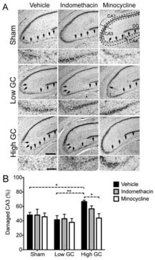
Fig. 3.
High GCs increase excitotoxic injury and this is reversed by minocycline. (A) Representative images of Nissl-stained sections following each treatment combination at 72 h post-KA. Damage was quantified in the CA3 region (magnified below each image). Hippocampal sub-regions are indicated with dashed lines. Arrows point to areas of neuron loss. Scale bars = 500 μm, (200 μm, magnification). (B) Quantification of CA3 lesioned area 72 h post-injury. High GCs significantly increased CA3 damage relative to low GCs and sham rats. This was reversed by minocycline, but not indomethacin treatment. Significance levels * p < 0.05 and ** p < 0.01 established by 2-way ANOVA followed by Holm-Sidak post hoc test. Significant 2-way ANOVA main effect of GC treatment (p < 0.01). Sham, vehicle n = 7; low GC, vehicle n = 12; high GC, vehicle n = 6; Sham, indomethacin n = 6; low GC, indomethacin n = 5; high GC, indomethacin n = 9; Sham, minocycline n = 7; low GC, minocycline n = 7; high GC, minocycline n = 6.

