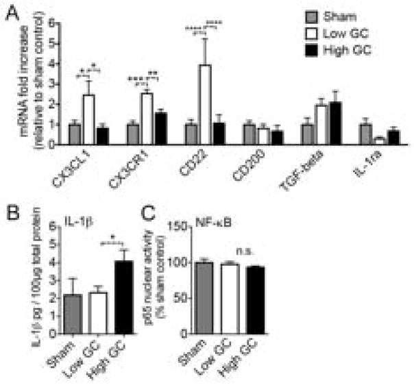
Fig. 5.
GCs suppress anti-inflammatory gene expression. Rats were treated with GCs and the hippocampus was dissected at the time when the injury would normally be given. n = 5–6 per group. (A) Anti-inflammatory gene expression in the uninjured hippocampus following each GC treatment, expressed as fold-increase relative to sham. Low GC animals had elevated CX3CL1, CX3CR1, and CD22 expression relative to sham or high GC treated animals (* p < 0.05, ** p < 0.01, *** p < 0.001, **** p < 0.0001). Significance established by 2-way ANOVA followed by Holm-Sidak post hoc test. (B) IL-1beta protein levels in the uninjured hippocampus following each GC treatment. High GC animals had higher IL-1beta levels than low GC animals (* p < 0.05). Significance established by two-way unpaired t test. (C) Quantification of EMSA autoradiographs for the p65 subunit of NF-kB in the uninjured hippocampus. Basal NF-kB activation was not affected by GC-treatments. (* p < 0.05, ** p < 0.01, **** p < 0.0001). Significance established by 2-way ANOVA followed by Holm-Sidak post hoc test.

