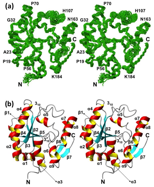
Fig. 2.
NMR structure of NP_344798.1. (a) Stereo view of a bundle of 20 NMR conformers representing the NMR structure. The chain ends and some sequence positions are identified to guide the eye. (b) Stereo ribbon representation of the conformer closest to the mean coordinates of the bundle shown in (a). β-strands are cyan, helices are red/yellow, and segments with non-regular secondary structure are grey. The chain ends and the regular secondary structures are identified. The figure was prepared using the program MOLMOL (Koradi et al. 1996).

