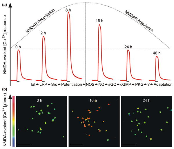
Fig. 1. HIV-1 Tat causes a biphasic change in NMDA-evoked increases in [Ca2+]i.

a, Stylized traces (
 ) depict time-dependent, biphasic changes in NMDA-evoked increases in [Ca2+]i following exposure to HIV-1 Tat for the time indicated above the trace. The previously reported (Krogh et al. 2014) sequence of events listed below the traces indicates that Tat activation of an LRP/Src kinase pathway resulted in NMDAR potentiation. Subsequently, NMDARs adapted following activation of the NOS/sGC/PKG signaling pathway. Identifying downstream effector(s) of PKG is the goal of this study. b, Representative pseudocolor images (scale bar = 100 μm) show peak response to NMDA (10μM, 30 s) in primary hippocampal neurons treated with Tat for the time indicated in the image panel. Images were scaled from 0 to 700 nM as indicated by the color bar on the y-axis.
