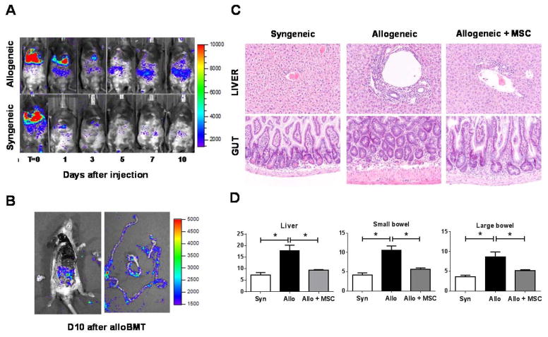
Figure 2. hMSCs migrate to abdominal organs and attenuate target-tissue GvHD cytotoxicity.
(A) Serial bioluminescence imaging (BLI) demonstrates that hMSCs home to the gastrointestinal tract in alloBMT mice. Data from one of three similar independent experiments is shown. (B) Post-mortem examination of intestines dissected from alloBMT mice that received hMSC infusions on D1 and D4. The mouse was euthanized and abdominal tissues dissected on D10. BLI was performed on dissected intestines. (C, D) Early reduction in proinflammatory T-cell expansion and cytokine induction as shown in Figure 1 associates with decreases in GvHD-associated histopathology in liver and bowel (C) and reduced GvHD organ histology scores (D). Organs harvested from five individual mice per transplant group were analyzed for GvHD severity at D10. Data are expressed as mean ± SEM and represent one of two similar experiments (n=5 mice per experimental group; * p < 0.01 and ** p < 0.05 for indicated comparisons by Mann-Whitney test).

