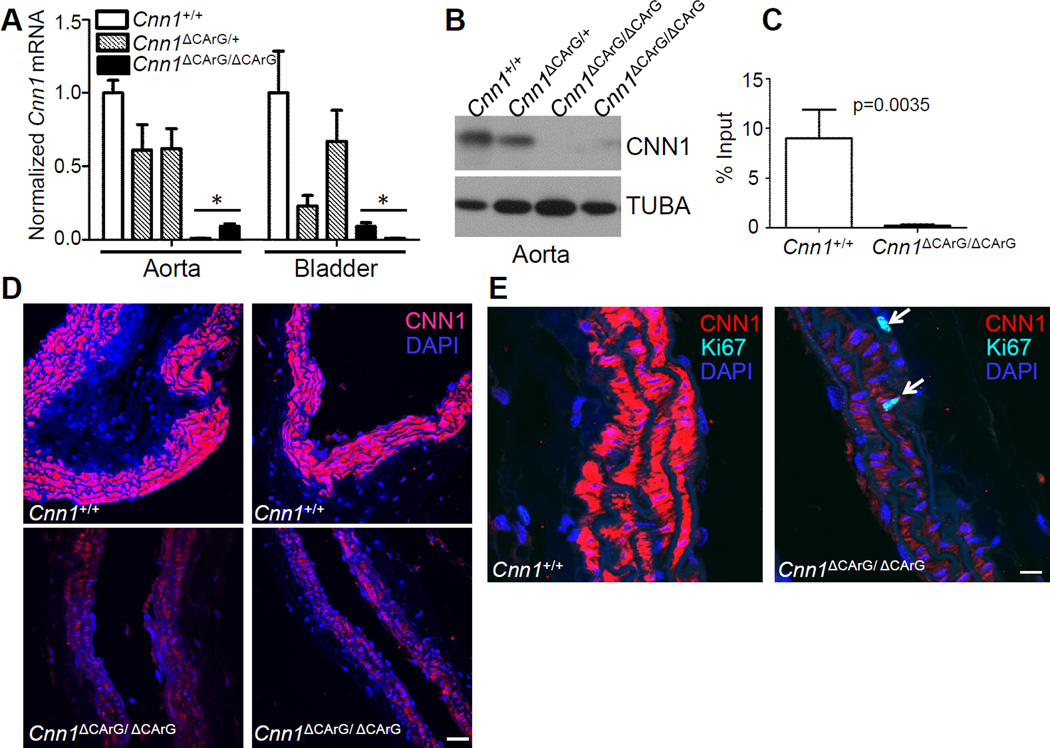
Figure 2. Expression of Cnn1 mRNA and CNN1 protein in F1 mice.
(A) Mouse aorta or bladder analyzed for expression of Cnn1 mRNA by quantitative RT-PCR with normalized Cnn1+/+ set to 1.0; other genotypes expressed relative to Cnn1+/+. (asterisks indicate p<0.01 compared to wildtype). See also Supplemental Figures II and III. (B) Expression of CNN1 protein in aorta by Western blotting. (C) In vivo chromatin immunoprecipitation assay of SRF binding to wildtype versus mutant intronic CArG box in bladder tissue. (D) Confocal immunofluorescence microscopy of CNN1 in wildtype versus mutant carotid arteries of two different founder lines. (E) SMC DNA synthesis (arrows) revealed with an antibody to Ki-67. Similar findings were seen in an independent founder line. Scale bars are 30 µm and 10 µm for panels D and E, respectively.

