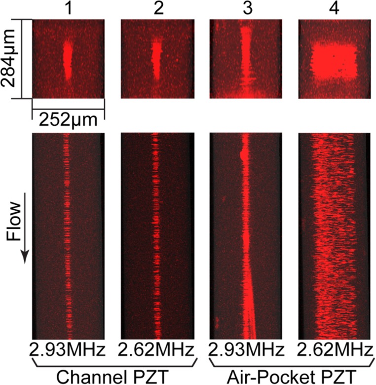
FIG. 5.
Stacked confocal fluorescence images of x-y planes (top) and x-z planes (bottom) of microchannels (W = 252 μm; H = 284 μm) at low flow rate. Cases 1–4 show image stacks obtained when Q = 20 μl/min. In Cases 1 and 2, the PZT transducer underneath the microchannel was activated at a driving frequency matching the resonant frequency either for width (Case 1) or for depth (Case 2) of the microchannel. Cases 3 and 4 are arranged in the same order as Cases 1 and 2 with regard to resonant conditions, but they were driven by a PZT transducer bonded underneath the air pocket.

