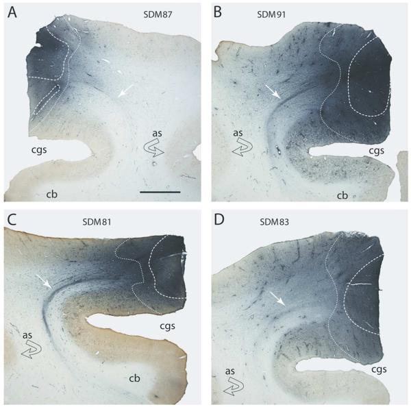
Figure 1.
Plate of low-power digital photomicrographs of immunohistochemically processed coronal tissue sections illustrating the fluorescein dextran (FD) injection site in the arm/hand region of M2 in F2P2 lesion cases SDM87 (A), SDM91 (B), SDM81 (C) and SDM83 (D). The dashed line represents the external boundary of the injection site core and the dotted line the external boundary of the injection site halo. The white arrows identify a coalesced descending labeled fiber bundle emerging from the FD injection site. The curved black arrow indicates the location of gray matter lining the depths of the arcuate spur. Abbreviations: as, spur of arcuate sulcus; cb, cingulum bundle; cgs, cingulate sulcus. The scale bar in A = 2mm and applies to all panels.

