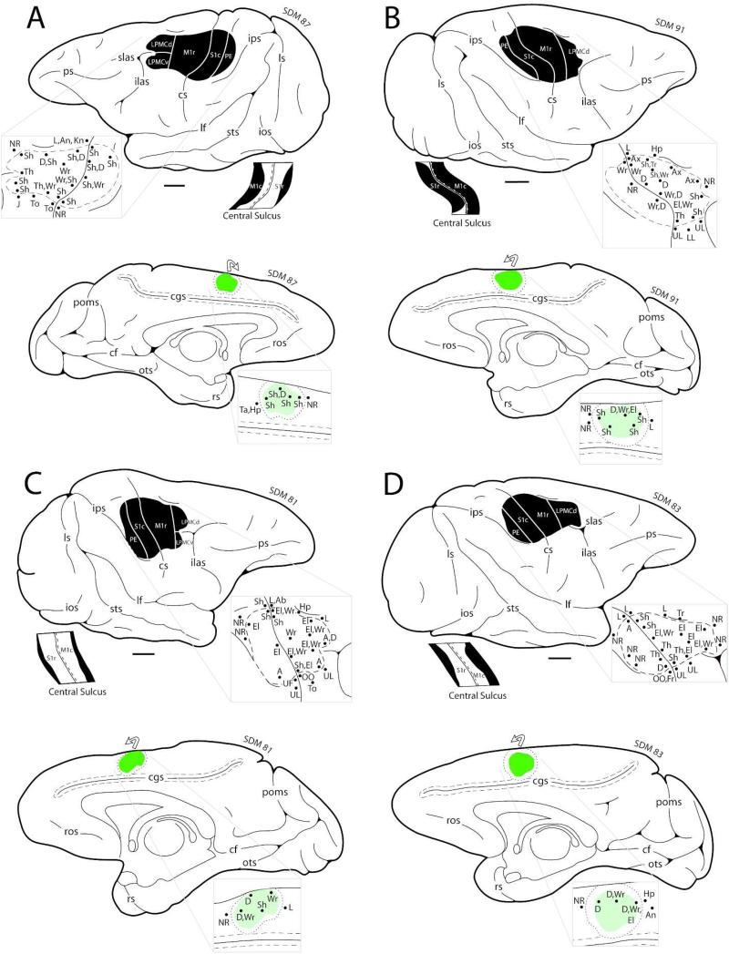
Figure 4.
Line drawings of the lateral (top) and medial (bottom) surfaces of the cerebral cortex in F2P2 lesion cases SDM87 (A), SDM91 (B), SDM81 (C) and SDM83 (D) depicting the locations of the lesion site (blackened area on lateral surface) and core of the fluorescein dextran (FD) injection site (green irregular shape on the medial surface). On the medial wall the dotted line around the injection site core represents the external boundary of the injection site halo. The curved arrow above the injection site indicates that some of the injection site halo was present on the dorsal convexity. On the lateral surface, the pullout depicts the physiological map of movement representation obtained with intracortical microstimulation used to localize the arm representations of the primary motor cortex (M1), lateral premotor cortex (LPMC), and primary somatosensory cortex (S1) prior to neurosurgical resection of the gray matter forming these cortical regions. Below the occipital lobe on the lateral surface drawing, is a flattened map showing cortex lining the rostral and caudal banks of the central sulcus. The black region represents the lesion site that extended into the sulcal cortex. The pullout on the medial surface is the physiological map of movement representation obtained with intracortical microstimulation to localize the arm representation of M2 prior to injection of the tract tracer FD. Scale bar = 5 mm and applies to the lateral surface, medial surface and flattened map of the cortex lining the central sulcus. For abbreviations see list.

