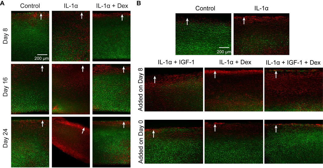
Figure 3.
A, Bovine chondrocyte viability in cartilage disks in response to 8, 16, or 24-day treatments. Treatment groups are control, IL-1α (1ng/mL), and IL-1α + Dex (100nM). Cells were fluorescently labeled with fluorescein diacetate (green, viable) and propidium iodide (red, non-viable). B, Bovine chondrocyte viability evaluated on Day 16 after treatments. Disks were treated with IL-1α (1 ng/ml) for the first 8 days, and switched to 1 pg/ml IL-1α between Day 8 and Day 16. IGF-1 (100 ng/ml), Dex (100 nM), or both were added either on Day 8 (middle panel) or Day 0 (bottom panel). White arrow: intact superficial surface of cartilage. Scale bar = 200 µm.

