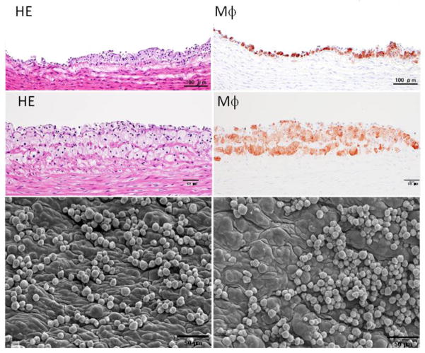
Fig. 3.
Early pathological changes of aortic surface of cholesterol-fed rabbits. Two representative lesions are shown (the top and middle panels) and were subjected to either hematoxylin and eosin (HE)staining (left) or immunohistochemical staining with RAM11 antibody against rabbit macrophage (Mϕ) (right). Aortic lesions are also visible under a scanning electron microscope (lower panels). Many monocytes either singly or in clusters adhere to the endothelial cells of the aorta.

