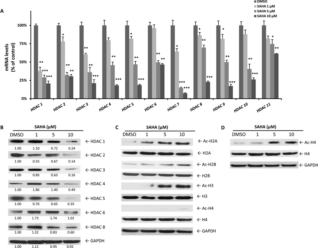
Figure 5.
SAHA reduces HDAC expression and enhances histone acetylation in leukaemic NK cells. (A) NKL cells were treated with SAHA (1, 5 and 10 µM) for 24 h and RNA was extracted for qRT-PCR analysis. Data are representative of two independent experiments performed in triplicate and normalized against 18S as a control gene. *P < 0.05, **P < 0.005, ***P < 0.0005 vs. dimethylsulphoxide (DMSO) control. (B) Western blot analysis was performed for HDACs after treatment with SAHA (1, 5 and 10 µM) for 24 h. Data were quantified by densitometry and ratios of HDAC/GAPDH are shown under each band. Results are representative of three independent experiments. (C) Western blot analysis was performed for acetyl-Histone after treatment with SAHA (1, 5 and 10 µM) for 24 h or (D) 48 h. Results are representative of three independent experiments. Equal loading of protein lysates was confirmed by re-probing membranes for GAPDH.

