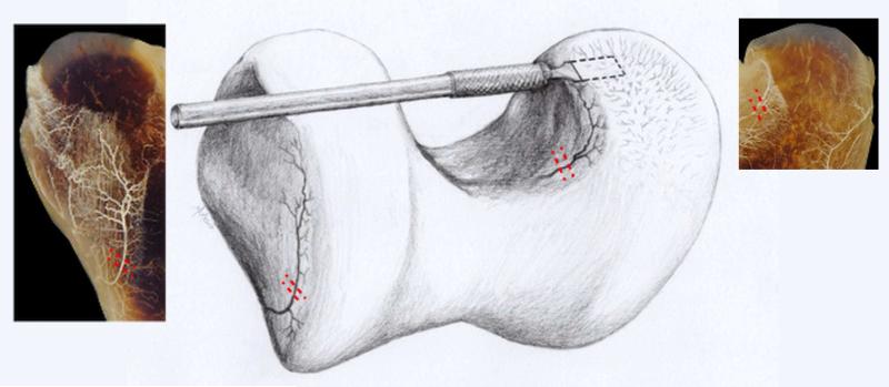
Figure 1.
Drawing demonstrating the surgical procedure used to interrupt the vascular supply to the distal femoral AECC. Dashed lines indicate location of incisions used to interrupt perichondrial vessels on the axial aspect of the medial femoral condyle (right) and lateral trochlear ridge (left). The incision extending into the epiphyseal cartilage is indicated by the beaver blade entering the medial femoral condyle. Perichondrial vessels interrupted in the axial aspect of the medial femoral condyle (right inset) and the abaxial aspect of the lateral trochlear ridge (left inset) are also shown in perfused, cleared specimens obtained from a 2-week-old goat.

