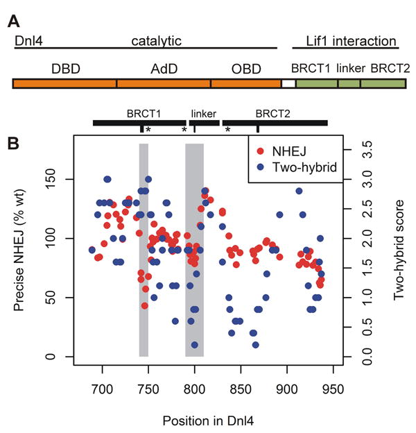
Figure 1. Separation of function in the Dnl4 BRCT mutation screen.

(A) Functional domains of Dnl4. DBD, DNA binding domain; AdD, adenylation domain; OBD, oligonucleotide binding domain; BRCT, BRCA1 C-terminal domain. (B) Panel showing precise NHEJ and Lif1 two-hybrid results for all point mutations tested in the Dnl4 BRCT mutation screen, plotted by their position in the Dnl4 protein. Each point represents a single mutant after application of a 5-point moving average over the position series to make regional patterns easier to visualize. Grey shading highlights two regions, 740-750 and 790-810, where different result patterns revealed a separation of function in the Dnl4 BRCT region. The spans of the two BRCT domains and inter-BRCT linker are denoted by black bars. Hash marks below denote the positions of mutations KTT(742:744)ATA, D800K, and GG(868:869)AA studied in further detail, and “*” indicates the position of an introduced stop codon.
