Figure 2.

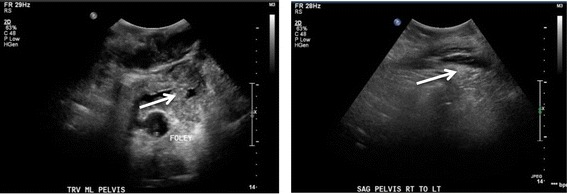
Ultrasonography of the upper pelvis, transverse (left) and sagittal (right) views. The heterogeneous mass (arrows) lies superficial to the bladder, which is decompressed with a Foley catheter.

Ultrasonography of the upper pelvis, transverse (left) and sagittal (right) views. The heterogeneous mass (arrows) lies superficial to the bladder, which is decompressed with a Foley catheter.