
Figure 3.
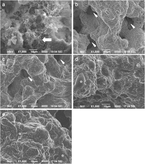
SEM images of biofilms prepared for observation using hydrophilic and hydrophobic ILs. (a): SEM images of platinum-coated S. mutans biofilms. Fibrous extracellular matrix-like structures (star) were partially observed. In some areas of the biofilm surface, the cells were separated from the biofilm (arrow). Images of biofilms prepared for observation using hydrophilic ILs (b) [Ch][Lac] and (c) [C2mim][AcO], and hydrophobic ILs (d) [C4mim][Tf2N] and (e) [P4,4,4,12][BF4]. The concentration of all ILs was 10%. Some hydrophobic ILs were repelled by the biofilm surfaces and pooled on them. An asterisk indicates the accumulated IL. Arrowheads indicate the dark gaps thought to be water channels. Scale bars = 10 μm.