
Figure 1.
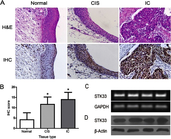
Expression of STK33 in human HSCC tissue and Fadu cells. (A) Representative imagines of STK33 IHC staining and matched H&E staining in hypopharyngeal normal and tumor tissues. a) Low STK33 expression in normal tissue; b) Moderate STK33 expression in CIS; and c) High STK33 expression in IC (Magnification × 400, Scale bars, 50 μm) (B) IHC score of STK33 expression in samples shown in A. Data were expressed as the mean ± SD. * P < 0.05. (C, D) The STK33 mRNA and protein were expressed abundantly in Fadu cells as determined by qRT-PCR and western blot.