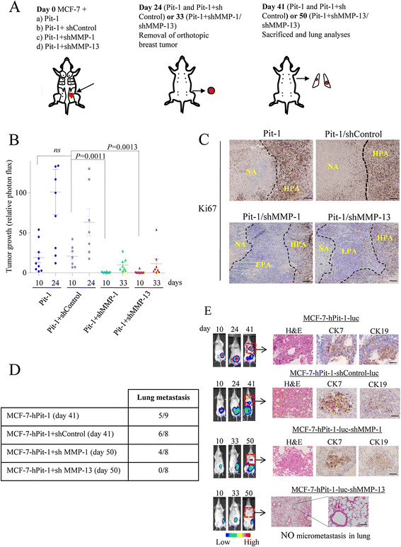
Figure 5.

Orthotopic injection of MCF-7 cells with Pit-1 overexpression and MMP-13 knockdown in SCID mice blocks metastasis to lung. (A) Schematic representation of experimental induction of metastasis. At day 0, SCID mice were injected into the mammary fat pad either with MCF-7 cells: (a) with Pit-1 overexpression (controls, n = 9), (b) with Pit-1 overexpression and shControl (controls, n = 8), (c) with Pit-1 overexpression and MMP-1 knockdown (n = 8), and (d) with Pit-1 overexpression and MMP-13 knockdown (n = 8). At day 24 (in mice with Pit-1 overexpression and shControl) or day 33 (in MMP-1 and MMP-13 knockdown mice), animals were anesthetized and breast tumors resected. Mice lived until day 41 (Pit-1 overexpression) or day 50 (Pit-1 plus MMP-1 or MMP-13 knockdown), and then were sacrificed and lungs removed for analysis. (B) Scatter plots of tumor growth in SCID mice at days 10 (all groups), 24 (mice with Pit-1 overexpression, Pit-1, and Pit-1 + shControl), and 33 (mice with Pit-1 overexpression and MMP-1 or MMP-13 knockdown), as described in A. Horizontal bars represent mean ± SEM. (C) ki67 immunostaining of tumors from mice injected with MCF-7 and Pit-1 overexpression (Pit-1), Pit-1 + shControl, Pit-1 + shMMP-1, or Pit-1 + shMMP-13. NA: necrotic area; HPA: high proliferative area; LPA: low proliferative area. Scale bar: 50 μm. (D) Five out of the nine mice with Pit-1 overexpression (day 41), six out of the eight with Pit-1 overexpression and shControl (day 41), and four out of the eight with Pit-1 overexpression and MMP-1 knockdown (day 50) developed lung metastasis, while none of the eight mice with MMP-13 knockdown (day 50) showed micrometastasis in lung. (E) Representative example of mice. Color indicates tumor cell luminescence. H&E staining and CK-7 and CK-19 immunopositivity in lung. Scale bar: 100 μm. CK, cytokeratin; H&E, hematoxylin and eosin; MMP-1, matrix metalloproteinase-1; MMP-13, matrix metalloproteinase-13; Pit-1, POU class 1 homeobox 1; SCID, severe combined immunodeficiency.
