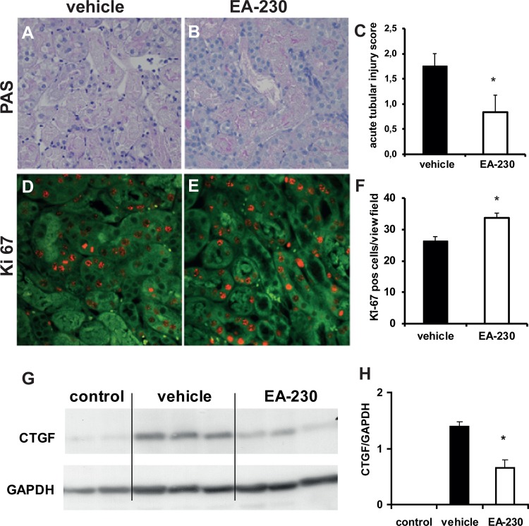
Figure 2. Acute tubular necrosis (A-C) two days after IRI affected ∼25% of tubuli in the vehicle treated mice and was less in the EA-230 treated mice (A-C, magnification 200 fold).
The number of Ki-67 positive tubular epithelial cells as marker of proliferation (red staining) was significantly higher in EA-230 treated kidneys two days after IRI (D-F, magnification 200fold, the autofluorescent kidney tissue appears green).

