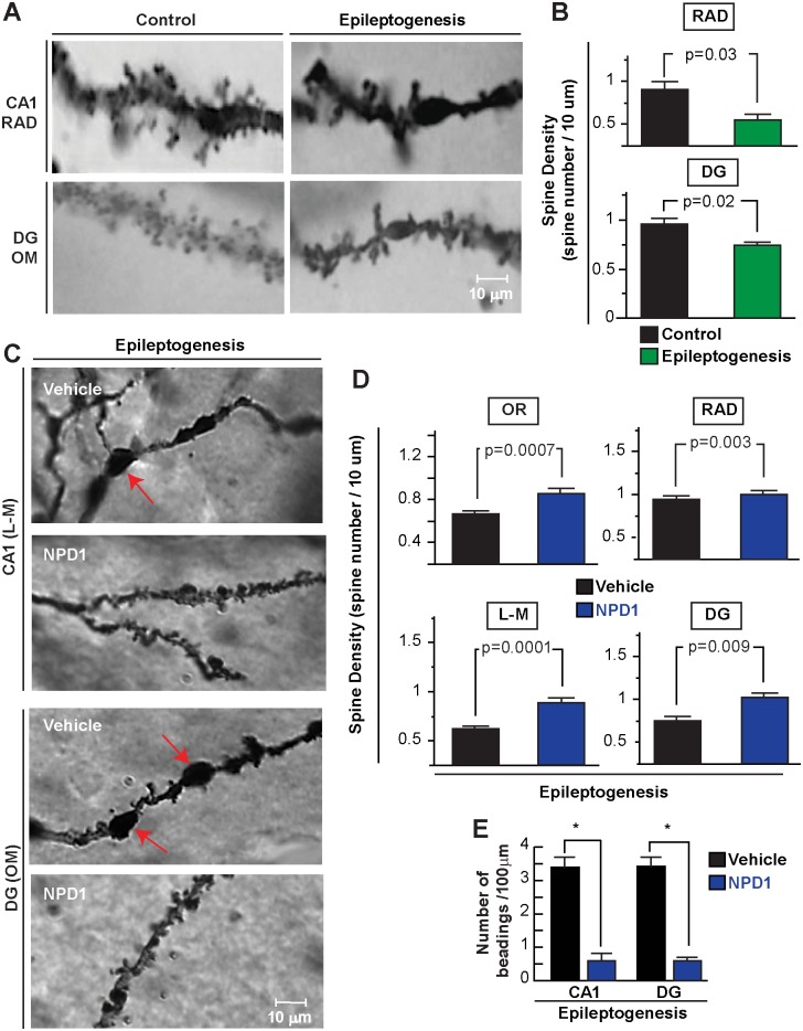
Figure 7. Neuroprotectin D1 induces sustained protection of dendritic spines during epileptogenesis.
A: Representative dendrites from control (naïve) and a mouse at 7 days after status epilepticus (SE) from CA1, stratum radiatum (RAD), dentate gyrus (DG) and outer molecular layer (OM). B: Note loss of number of dendritic spines as a consequence of status epilepticus (PSE, n = 3) compared with control (n = 3). C: High-power light magnification of dendrite profiles of CA1 and dentate gyrus (DG) regions showing dendritic beadings in vehicle-treated mice (n = 4) (arrows) and lower dendritic spine profiles compared with NPD1-treated mice (n = 4) in epileptogenesis D: NPD1-treated mice show a higher number of dendritic spines in the hippocampal layers than vehicle. E: Number of dendritic beadings per dendrites is reduced in NPD1-treated mice compared with vehicle. Bars indicate means, and error bars represent S.E.M. p = p value. *: p<0.05, two sample t-test.

