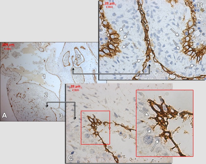
Figure 7. Human embryonic heart (56 days), CD 31 immune labeling.
Immune labeling with CD31 antibodies of a 56 days human embryonic heart, oblique-sagittal cut. A general view is presented in (A) and detailed in (B) and (C) (corresponding fields are indicated by black connectors). An area in (B) is magnified (inset). The white arrows indicate endocardial tip cells projecting moniliform filopodia within the subendocardial stroma. 1.atrioventricular canal; 2.atrioventricular cushion; 3.ventricle; 4.aortic cushion; 5.aortic sinus.

