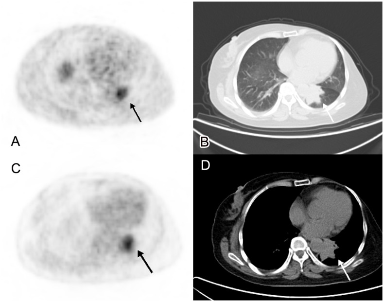
Fig 1. A 58-year-old female breast cancer patient.

We detected a mass with irregular margin in left lung (B,D. CT imaging), with a maximum diameter of 4.5cm. It was difficult to differentiate it between metastasis and secondary primary lung cancer in CT imaging. The tumor presented with high uptake of both 18F-FES and 18F-FDG PET, SUVmax was 6.3 and 5.5 respectively. It suggested that it was a metastasis (A. 18F-FES PET/CT, C. 18F-FDG PET/CT). After operation, it was confirmed to be an ER positive metastasis from breast cancer.
