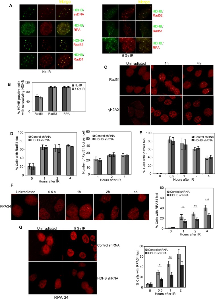
Figure 3. HDHB depletion impairs RPA late-stage foci formation after IR.
(A) U2OS cells transiently expressing GFP-HDHB were stained with different antibodies and observed by immunofluorescence. To visualize ssDNA, cells were grown in BrdU for 24 hours, fixed and stained with anti-BrdU antibody. Left, no IR; Right, cells were fixed at 2 h after 5 Gy IR treatment. (B) Percentage of HDHB-positive cells with Rad51, Rad52 or RPA foci colocalizing with HDHB. 500 cells in three experiments were counted. The mean values ± s.d. are plotted. (C) Example of 5 Gy IR-induced Rad51 and γH2AX foci in HCT116 cells at 1 h or 4 h after irradiation. (D) Percentage of cells with Rad51 foci after IR. Right panel is the mean number of Rad51 foci per cell after IR. Total 1000 cells in three experiments were counted. The mean values ± s.d. are plotted. (E) Percentage of cells with γH2AX foci after IR. (F) Left, example of RPA34 foci in HCT116 cells at 0.5 h, 1 h, 2 h, 4 h after 5 Gy IR. Right, percentage of cells with RPA34 foci after IR. *P<0.05, **P<0.05, ***P<0.05, Student t-test. (G) Left, example of RPA34 foci in U2OS cells transfected with control shRNA or HDHB shRNA at one hour after 5 Gy IR. Right, percentage of cells with RPA34 foci after 5 Gy IR at different time points. *P<0.05, **P<0.05, Student t-test.

