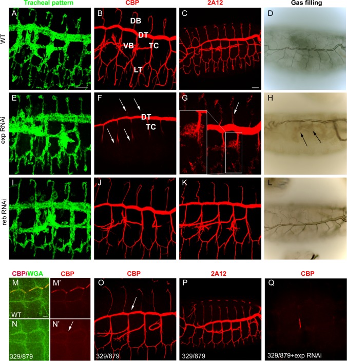
Figure 2. Tracheal defects in exp and reb loss-of-function.
All images are projections of confocal sections except D, H, and L, which are bright field images. All images show tracheal metameres of embryos at stage 13 (M,N), larval stage (D,H,L), or stage 15 (rest of panels). In WT embryos (A), chitin (B) and chitin-associated proteins (C) accumulate in the lumen of all tracheal branches, and by the end of embryogenesis the trachea fill with air (D). The down-regulation of reb does not generate detectable defects (I-L). In contrast, exp down-regulation prevents luminal accumulation of chitin and associated proteins in all branches (arrows in F,G), except the DT and part of TC, while the pattern of branching is normal (E). Later, only the DT is filled with air (arrows in H). In the absence of reb, chitin deposition in the DT is delayed (arrow in N’, compare to M’). Here WGA allows visualisation of the apical region of the trachea (M,N). However, at later stages, chitin (O) and associated proteins (P) are present (with slightly lower DT levels, arrow in O). When exp is down-regulated in the deficiency combination, no chitin accumulates in the trachea (Q). Scale bars A,C 25 μm, M 7.5 μm.

