Image 1.
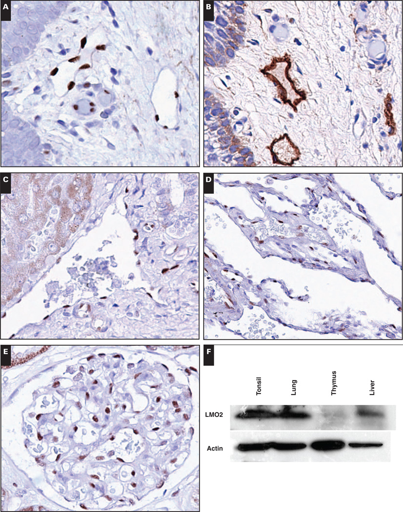
Native tissue vasculature, including lymphatic vasculature, is generally LMO2+. A, Tonsil shows LMO2+ blood vessels and lymphatic vessels (×400). B, Tonsil stained with lymphatic marker D2-40 to highlight lymphatic vessels (×400). C, Liver shows LMO2+ portal arteries and veins and LMO2− sinusoids (×400). D and E, Lung (D, ×400) and kidney (E, ×400) have LMO2+ vasculature. F, Expression of LMO2 in native tissues by immunoblotting. Actin serves as a loading control.

