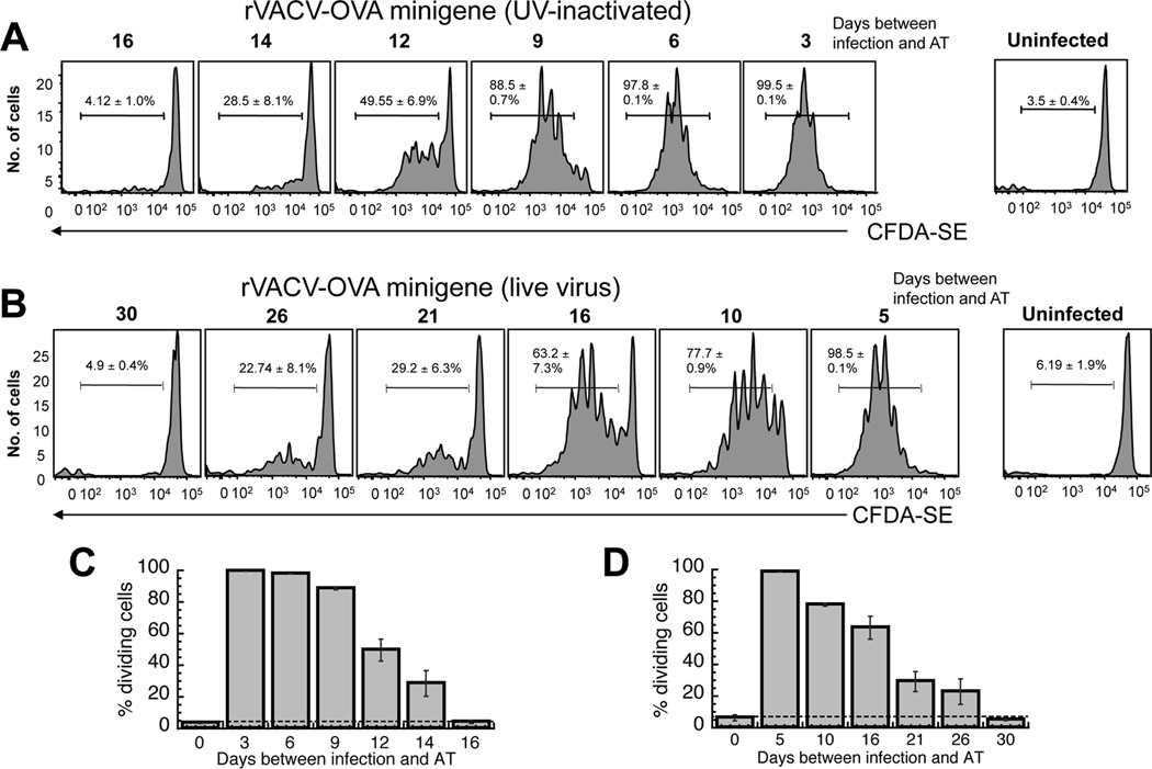
Figure 3. The requirement for presentation by rVACV infected cells in prolonged antigen presentation.
C57BL/6 mice were infected with non-replicating (A) or replicating (B) rVACV-OVA MG at times indicated. Virus replication was blocked with a 20 minute treatment with UVA/psoralen. OT-1.SJL T cells were labeled with CFDA-SE and transferred i.v. into previously infected mice. 3 days after transfer, spleens were removed and analyzed for CFDA-SE dilution. Representative histograms display gated CD45.1+ TCD8+ from individual mice of replicate experiments using 6 mice per condition. Gates represent the percentage of CD45.1+ TCD8+ cells that proliferated. (C, D) Graphical representation of the percentage of replicating OT-1.SJL TCD8+ cells at each time point after VACV infection in A and B respectively. Error bars represent the standard error.

