
Figure 5.
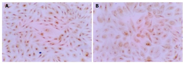
Positive expression with brown dye occurred in cytoplast of MMP2 protein of GBC-SD cells (immunohistochemistry SABC method, ×100). A: The brown dye of MMP2 was shown positively in most cells of the control group. B: In the experiment group with treatment of NCTD (5 μg/mL) for 48 h, the positive cells of MMP2 expression decreased significantly and the dye in the cytoplast became light.