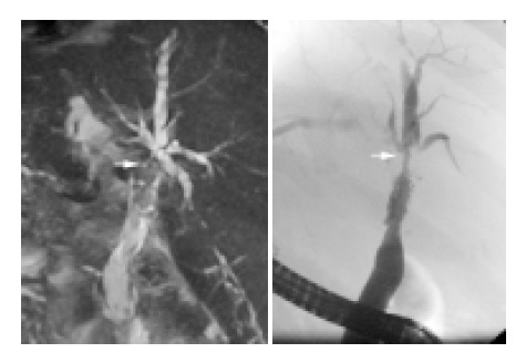
Figure 2.

Comparison of ERC and MRC in posttransplant biliary stenosis. ITBL type III with hilar stenosis (arrow) and multiple peripheral duct stenoses. Left: MRC, right: ERC. The dominant hilar stenosis (arrow) is seen with both methods. The peripheral stenoses are better seen in ERC.
