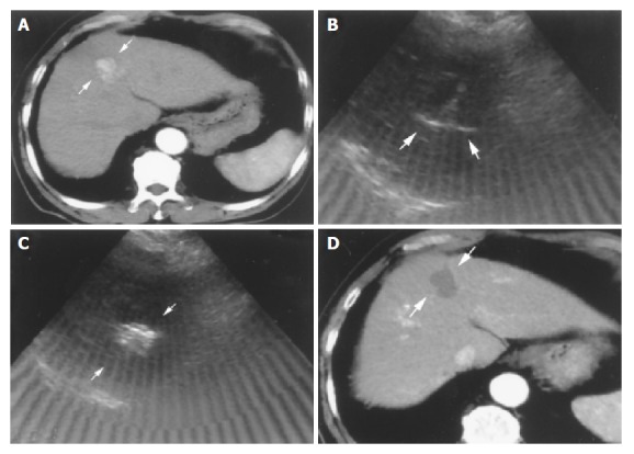
Figure 1.

Successful RFA in an 82-year-old male patient with HCC. A: Tri-phase CT scan showed an enhanced nodule in hepatic arterial phase about 2.8 cm in size before RFA (arrows); B: Radiofrequency ablation was performed in one session. The needle was introduced into the lower part of the tumor first with fully expanded tines under real-time ultrasound-guidance (arrows); C: Complete ablation after 2 ablative courses within 37 min was done. The tumor showed hyperechoic signals after RFA under ultrasound (arrows); D: Tri-phase CT scan showed complete necrosis of this tumor without enhancement in hepatic arterial phase one month after RFA (arrows).
