Figure 2.

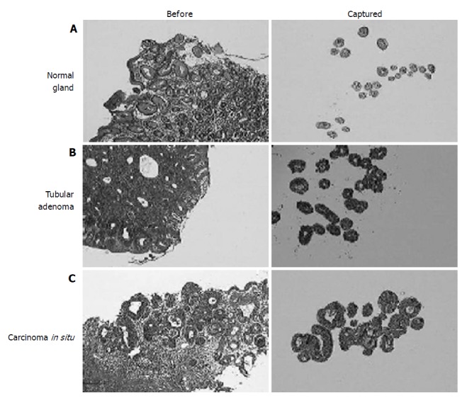
Histologic view (left) and microdissected cells (right) of the normal gastric mucosa. (A) adenoma; (B) and carcinoma in situ; (C) (HE stain. ×20). Compared to adenomas, carcinoma in situ had only mild glandular complexity.

Histologic view (left) and microdissected cells (right) of the normal gastric mucosa. (A) adenoma; (B) and carcinoma in situ; (C) (HE stain. ×20). Compared to adenomas, carcinoma in situ had only mild glandular complexity.