
Figure 5.
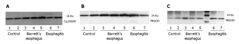
Representative Western analysis of tissue lysates (22 μg/lane) from control subjects (normal esophageal mucosa) and patients with esophagitis and Barrett’s esophagus. A: Western blot analysis of Cu,ZnSOD expression in esophageal biopsies demonstrated an increase of Cu,ZnSOD expression in Barrett’s esophagus biopsies (lanes 3-5) compared to control subjects (lanes 1,2) and patients with esophagitis (lanes 6,7); B: A band at 24 ku corresponding to monomeric MnSOD was observed in all groups evaluated. Increased staining for MnSOD was observed in Barrett’s esophagus (lanes 3-5) and esophagitis (lanes 6,7) compared to control patients (lanes 1,2); C: Immunoprecipitated tyrosine-nitrated proteins and subsequent incubation with anti-MnSOD demonstrated that MnSOD nitrated is increased in patients with esophagitis (lanes 6,7) and Barrett’s esophagus (lanes 3-5) compared to normal subjects (lanes 1,2).