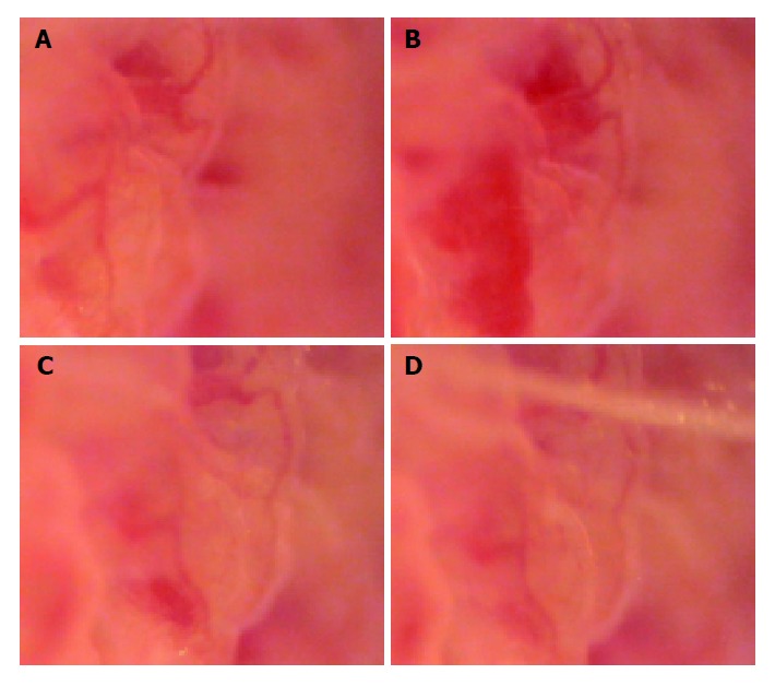
Figure 3.

Changes of microcirculation of jejunal mucosa. A: Photograph of microcirculation of jejunal mucous membrane, 6 s before the load of rabbit’s brainstem hemorrhage (×185); B: Photograph of jejunal mucous membrane, 10 min 26 s after the load of rabbit’s brainstem hemorrhage; it showed the local congestion of villus mucosa (×185); C: Photograph of jejunal mucous membrane, 30 min 32 s after the load of rabbit’s brainstem hemorrhage; it showed that the local congestion of villus mucosa had relieved (×185); D: Photograph of jejunal mucous membrane, 40 min 46 s after the load of rabbit’s brainstem hemorrhage; it showed that the local congestion of villus mucosa had ameliorated obviously (×185).
