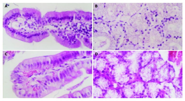
Figure 6.

HE stain of duodenal structure of rabbits. A: HE stain of normal rabbit’s duodenal villus. The minute blood vessel had not dilated and red blood cells had not leaked out from capillary lumens; B: HE stain of normal rabbit’s mucous glands of duodenum. The minute blood vessel had not dilated; C: HE stain of the rabbit’s duodenal villus, which were 48 h after brainstem hemorrhage. The capillaries had obviously dilated and red blood cells had leaked out from capillary lumens; D: HE stain of the rabbit’s mucous glands of duodenum, which were 48 h after brainstem hemorrhage. The capillaries had obviously dilated and red blood cells had leaked out from capillary lumens.
