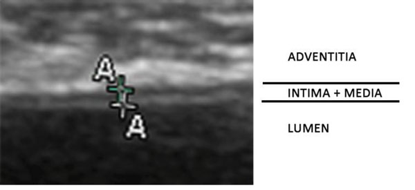
Figure 3.

A typical double line pattern ultrasound image of a normal abdominal aortic artery wall of a monkey. The distance between the centers of the two crosses represents the distance between the inner and the outer echogenic lines and corresponds to the ultrasound image of intima-media thickness.
