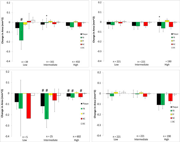
Figure 6.

Changes in VH-IVUS derived plaque component area in low-, intermediate-, and high WSS sectors in patient 1 for models with (top) and without (bottom) side-branches. Data are further divided into WSS (left) and relative WSS (right), n refers to the number of plaque sectors in each WSS category (note, the number of sectors of any individual plaque component in a specific WSS category may differ from this as not each sector contains every plaque component). *P < 0.05 comparing the model with side-branches to the model without side-branches (top plot to bottom plot) waveform; # P < 0.05 comparing WSS to relative WSS differences (left plot to right plot)
