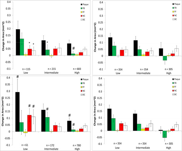
Figure 8.

Changes in VH-IVUS derived plaque component area in low-, intermediate-, and high WSS sectors in patient 3 for models with (top) and without (bottom) side-branches. Data are further divided into WSS (left) and relative WSS (right), see figure 5 for figure detail.
