Figure 1.

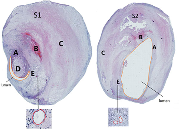
Two samples of microscopic slices of plaque stained using H&E. (Left: S1; Right: S2). A: Fibrous cap; B: Fresh intraplaque hemorrhage; C: Vessel; D: Lipid core; E: Neovessels.

Two samples of microscopic slices of plaque stained using H&E. (Left: S1; Right: S2). A: Fibrous cap; B: Fresh intraplaque hemorrhage; C: Vessel; D: Lipid core; E: Neovessels.