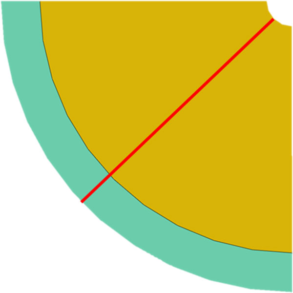
Figure 3.

Idealised concentric quarter model of a diseased femoral artery with an initial stenosis obstructing 90% of the luminal diameter. The yellow section represents the diseased tissue and the green section represents the healthy tissue. The red line indicates the line along which data was extracted from the model.
