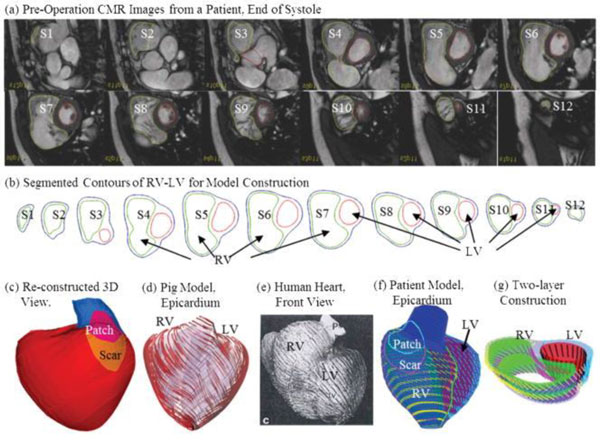
Figure 6.

A model construction procedures using cardiac MR imaging for CFD: a) cardiac MR images for a subject, b) contouring for the ventricles, c) reconstructed 3D chambers, d) a pig model showing myocardial fiber orientation, e) a human heart with fiber orientation, f) CFD model incorporating myocardial fiber orientation, and 3) two-layer model [62]
