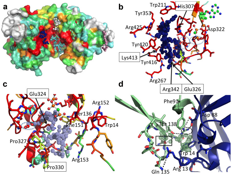
Figure 6. Identification of surface cavities in MakMvan.
(A) Surface representation of free MakMvan coloured according to residue conservation (consistent with the multiple sequence alignment shown in Supplementary Fig. S1). Cavities identified with the software fpocket36 and mentioned in the main text are represented as dotted spheres (pocket 1: blue; pocket 2: cyan; pocket 3: white; pocket 4: light green; pocket 5: light pink). (B) Close view of the residues lining pocket 1 coloured as in panel A. Residue Asp305 is represented as ball-and-stick and highlighted by a dashed blue line; the γ-phosphate of AppCp (ball-and-stick representation) is highlighted by a blue line. (C) Detailed view of residues delimiting pocket 2, coloured as in panel A; Asp305 is represented as ball-and-stick and circled by a dashed line. (D) Close view of the cavity located at the interface between the intermediate (green) and the N-terminal cap domain (blue); Asn137 is highlighted by a rectangle and Arg152 by a dashed ellipse. Hydrogen bonds are represented as dashed lines. For panels B–D, important residues are shown as sticks with oxygen atoms in red, nitrogen in blue, sulfur in yellow, phosphorus in orange and carbon in green (nucleotide) or according to sequence conservation (protein). Panels C–D refer to the structure of MakMvan in complex with AppCp. Figure prepared with PyMOL (http://www.pymol.org).

