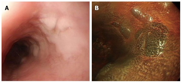
Figure 6.

Glycogenic acanthosis. A: Esophagogastroduodenoscopy reveals multiple, uniformly sized, oval or round glycogenic acanthosis usually < 1 cm, involving otherwise normal esophageal mucosa; B: In chromoscopy with iodine spray, glycogenic acanthosis is recognized as slightly elevated iodine-positive, brownish areas.
