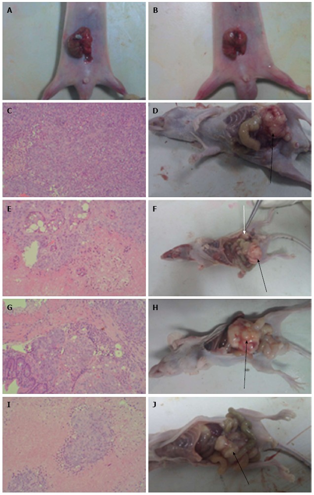
Figure 1.

Pathology of primary colon cancer tissues (HE staining) and gross anatomy of nude mice implanted with human colon cancer HCT-116 cells. A, B: Tumor pieces were transplanted into the cecum of nude mice by purse-string suture; C, E, G and I: HE staining of colon cancer tissues from mice in the CON, WCA, 5-FU and WCA + 5-FU groups (magnification × 100); D, F, H and J: Gross anatomy of mice in the CON, WCA, 5-FU and WCA + 5-FU groups. Black arrows indicate orthotopic tumors, and white arrows indicate the abdominal wall tumor. WCA: Weichang’an; 5-FU: 5-fluorouracil; CON: Control.
