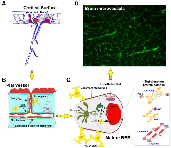
Figure 1.
Structure and location of brain microvessels. (A) Gross view of brain microvessels crossing through brain parenchyma, (B and C) Schematics of the inner view of the brain microvessels lined with closely associated endothelial cells together with pericytes and astrocytic foot processes, (D) FITC albumin stained brain section showing the overall network of brain microcapillaries. Note: VR (Virchow-Robin space).

