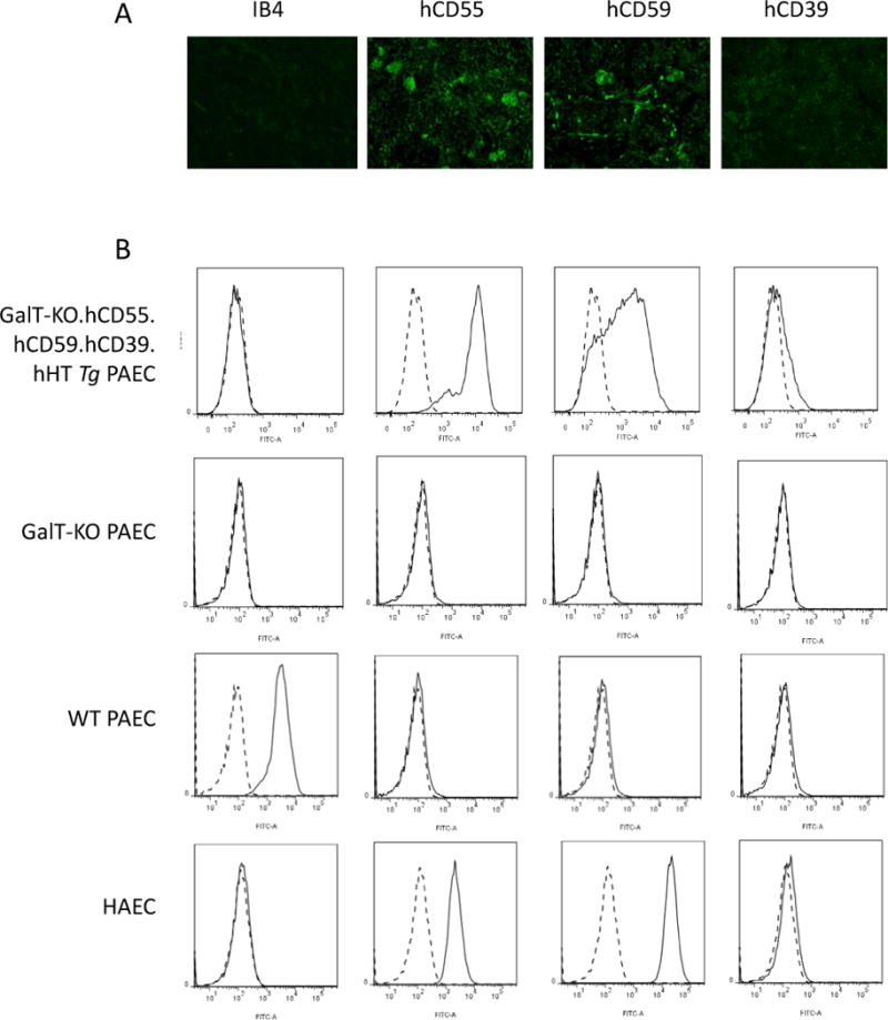
Figure 1.

Transgene expression and knock-out in pig cells and tissue. The expression of Gal epitope (IB4), hCD55, hCD59 and hCD39 was assessed by immunofluorescence staining on GalT-KO.hCD55.hCD59.hCD39.hHTTg kidney (1A) and by FACS on human (HAEC) and porcine aortic endothelial cells (PAEC) (1B): first row: GalT-KO.hCD55.hCD59.hCD39.hHTTg PAEC (donor from Control group and groups #1–3), second row: GalT-KO PAEC (donor from group #4), third row: PAEC WT and fourth row: HAEC. Histograms show negative control (cells without Ab, dashed line) and endothelial cells with Ab (plain line).
