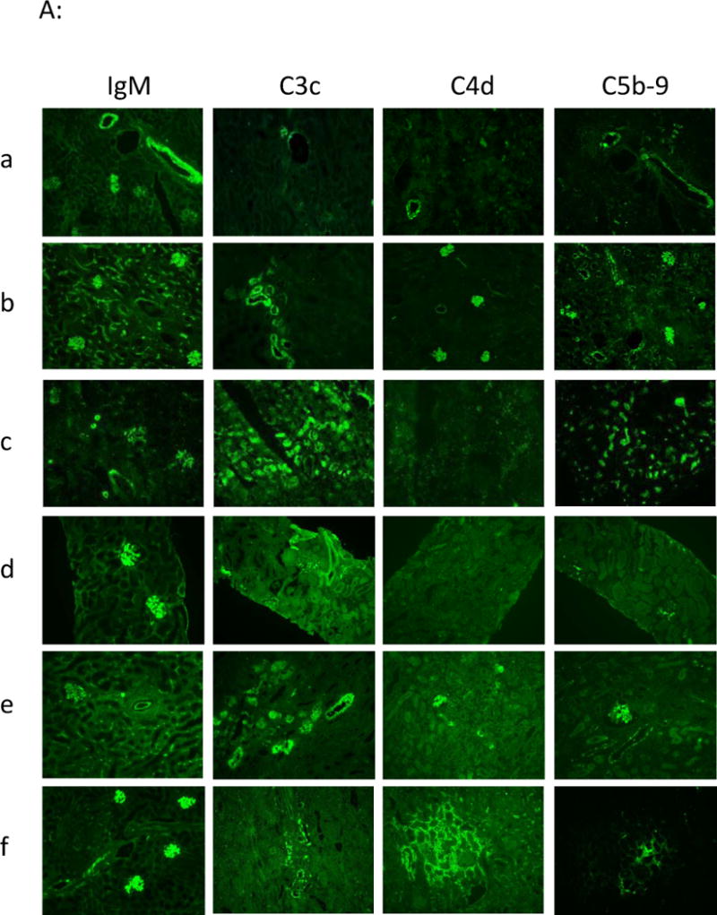
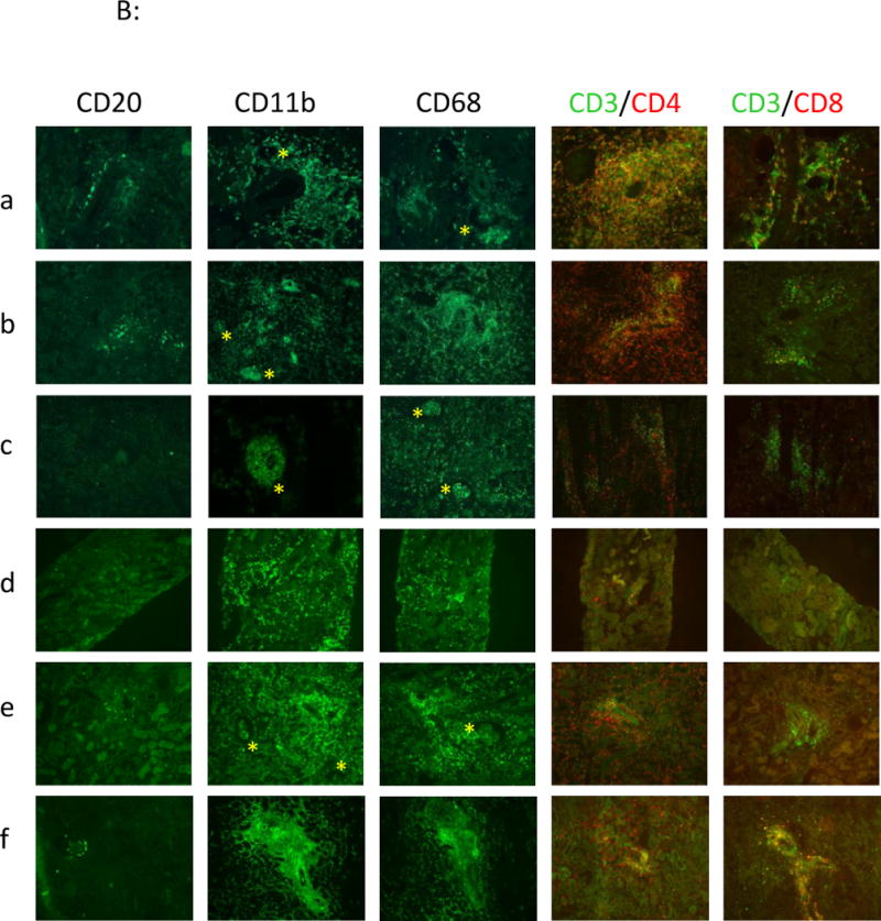
Figure 5.


Immuno-histofluorescence analyses. IgM, C3c, C4d, C5b-9 staining (4A) and cellular staining: B cells (CD20), monocytes/macrophages (CD11b, CD68): yellow star is emphasizing the presence of monocytes/macrophages in glomerular capillaries, CD4 and CD8 T cells (4B), were performed in frozen kidney biopsies from recipients a: control group (recipient #V9910C) at rejection at d3, b: from group#1 at rejection at d15 (recipient #V893AA), c: from group# 2 at rejection at d9 (recipient #PA997C) and d & e: from group#3 (recipient #PA936G) respectively at d4 in functioning graft (d) and at rejection at d12 (e), and f: from group#4 (recipient #V906F) at rejection at d11.
