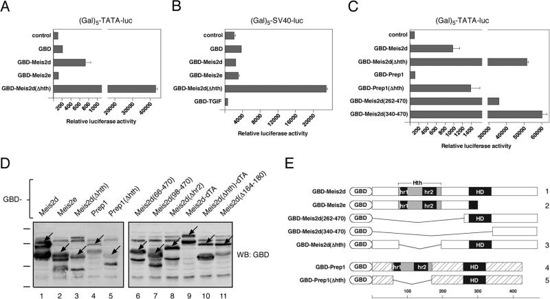
Fig. 1.
Meis2 contains a carboxyl-terminal activation domain. HepG2 cells were transfected with the indicated GBD fusion constructs and the (Gal)5-TATA luciferase reporter (A), or the (Gal)5-SV40 reporter (B). Luciferase activity was assayed after 48 hours and is presented as the mean + s.d. of duplicate transfections (arbitrary units). C) a series of Meis2 and Prep1 deletion constructs fused to the GBD were assayed as in A. D) The relative expression of the indicated GBD-fusions was analyzed by western blot with a GBD antibody. The specific full length bands are indicated by arrows. Numbers below each lane correspond the numbered constructs in Figures 1E and 4F. The positions of molecular weight markers (95, 72, 55 and 43kD) are shown to the left. E) GBD expression constructs are shown schematically. Hth: homothorax homology domain, hr1 and hr2: homology regions 1 and 2, HD: homeodomain. Scale below shows amino acid numbers.

