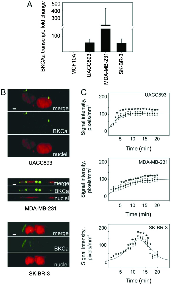
Figure 1.
BKCa channels regulate the transmembrane potential in breast cancer cells. (A) Breast cancer cells displayed higher levels of BKCa transcripts compared to benign mammary epithelial cells. Total RNA from MCF10A benign mammary epithelial cells and UACC893, MDA-MB-231 and SK-BR-3 beast cancer cells was assayed for BKCa transcript levels using quantitative RT-PCR. Statistically significant differences vs. the MCF10A cells. N=3, P≤0.05. (B) BKCa channel proteins (green) were detected in UACC893 (upper panels), MDA-MB-231 (middle panels) and SK-BR-3 (lower panels) cells using fluorescent immunocytochemistry. Nuclei were counterstained with propidium iodide (red) and the image series were acquired using Zeiss 510 laser confocal microscope. A series was reconstructed to generate three dimensional images rotated to present the lateral sides of the cells with apical surfaces facing atop of their respective image. MDA-MB-231 cells did not possess basal-apical differentiation and appear flat in the images. Experiments were repeated at least three times for each cell line. (C) IbTX differentially modulates transmembrane potential. UACC893 (upper panel), MDA-MB-231 (middle panel) and SK-BR-3 (lower panel) cells pre-loaded with 2 μmol/l DiBac4(3) in membrane potential buffer and supplemented with 10 nmol/l IbTX were observed using a Zeiss 510 laser confocal microscope for 20 min with images captured every minute. Average signal intensities from 5 to 7 regions of interest were calculated and plotted. Increased signal intensity signifies depolarization due to IbTX inhibition of BKCa channels by IbTX. Significant differences vs. start of experiment (1 min). N=3. IbTX, iberiotoxin.

