Figure 1.
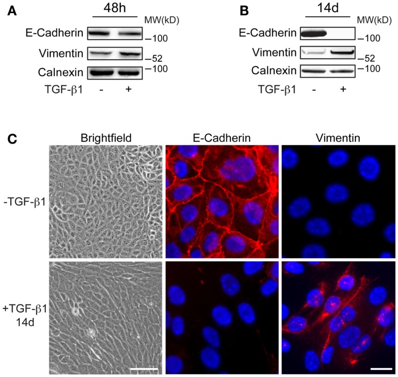
Characteristics of long-term EMT cells. (A,B) Western blot analysis showing more prominent changes in E-cadherin and vimentin expression in EpRas tumor cells after 14 days (B) compared to 48 h (A) of TGF-β1 exposure. Calnexin was used as a internal loading control. (C) Brightfield and immunofluorescence images showing that EpRas cells exposed to TGF-β1 for 14 days acquire characteristic features of EMT including an elongated, fibroblast-like morphology (left panels), loss of E-cadherin at cell–cell junctions (middle panels), and cytoplasmic staining of vimentin (right panels). Scale bars: 20 μm (left panels); 5 μm (middle and right panels).

